PRP 치료 그림
25 동료 심사 연구의 그림
Treatment options for androgenetic alopecia range from FDA-approved pharmacological agents (minoxidil and finasteride) to emerging therapies including low-level laser therapy, platelet-rich plasma, and hair transplantation. Efficacy, side effect profiles, and patient compliance vary substantially across modalities.
Treatment options for androgenetic alopecia: Efficacy, side effects, compliance, financial considerations, and …
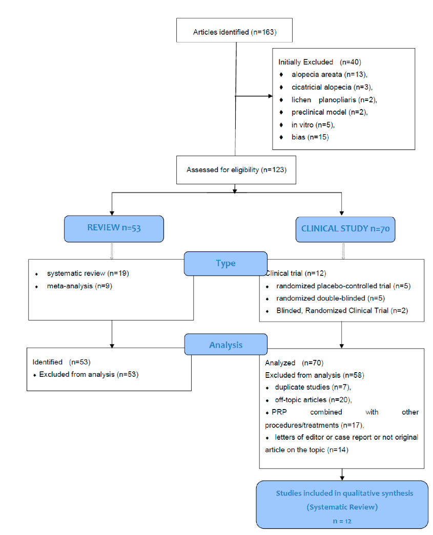
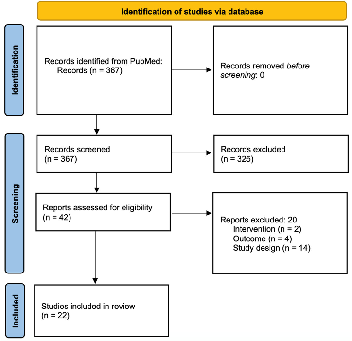
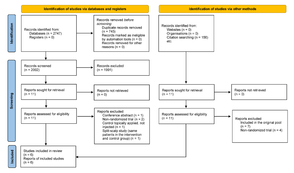
The CONSORT (Consolidated Standards of Reporting Trials) flow diagram documents the systematic review process from identification through screening, eligibility assessment, and final inclusion of studies evaluating PRP for androgenetic alopecia.
Systematic Review of Platelet-Rich Plasma Use in Androgenetic Alopecia Compared with Minoxidil®, …
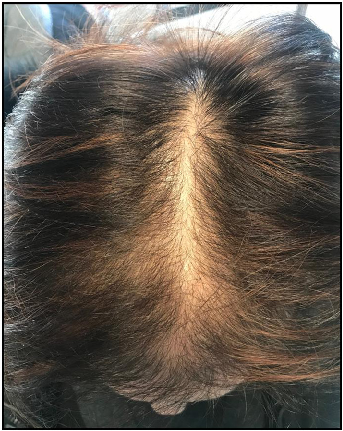
A clinical photograph demonstrating female pattern hair loss, showing characteristic diffuse thinning across the crown and mid-scalp. FPHL affects women with varying degrees of severity and may lead to significant psychological distress.
Female pattern hair loss: A clinical, pathophysiologic, and therapeutic review.
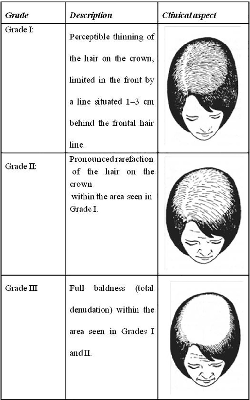
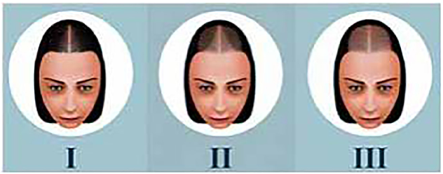
The Ludwig scale classifies female pattern hair loss into three progressive stages, ranging from minimal thinning at the crown (Grade I) to extensive hair loss across the top of the scalp (Grade III). This grading system remains one of the most widely used clinical tools for assessing FPHL severity.
Female pattern hair loss: A clinical, pathophysiologic, and therapeutic review.
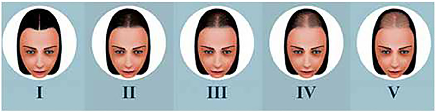
Sinclair's classification divides midline pattern alopecia into four intensity levels, progressing from a normal-appearing scalp to increasingly visible widening of the central part. The scale, introduced by Sinclair et al. (2005), provides a practical visual reference for clinicians assessing hair loss severity.
Female pattern hair loss: A clinical, pathophysiologic, and therapeutic review.
Olsen's classification system highlights the characteristic triangular or Christmas-tree pattern of frontovertical alopecia seen in female pattern hair loss. The accentuation of thinning at the frontal midline distinguishes this pattern from the more diffuse Ludwig classification.
Female pattern hair loss: A clinical, pathophysiologic, and therapeutic review.
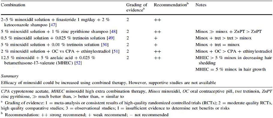
A summary of clinical evidence supporting combination therapy with topical minoxidil for androgenetic alopecia in both men and women. The data, compiled by Varothai and Bergfeld (2014), indicates that multi-modal treatment approaches may offer improved outcomes compared to monotherapy.
Female pattern hair loss: A clinical, pathophysiologic, and therapeutic review.
PRISMA flowchart detailing the systematic literature search and screening process for studies on microneedling in hair loss disorders. The diagram tracks records from initial identification through screening, eligibility assessment, and final inclusion.
Microneedling and Its Use in Hair Loss Disorders: A Systematic Review.
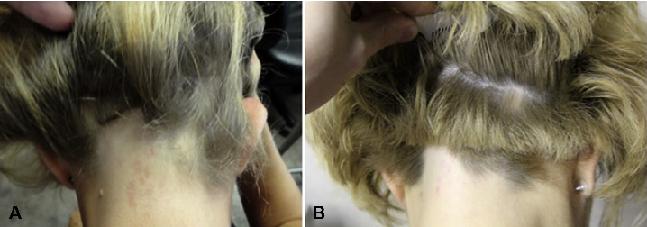
Clinical photographs showing hair regrowth in a patient with corticosteroid-resistant ophiasis-type alopecia areata before and 3 months after platelet-rich plasma (PRP) injection, with occipital hair loss markedly improved.
Successful treatment of corticosteroid-resistant ophiasis-type alopecia areata (AA) with platelet-rich plasma (PRP).
Alopecia and platelet-derived therapies.
Alopecia and platelet-derived therapies.
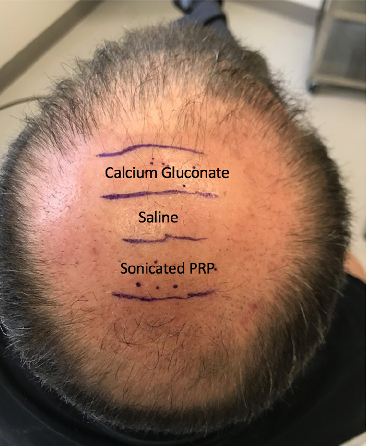
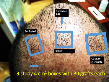
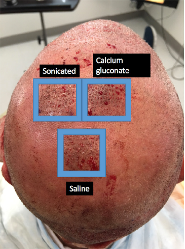
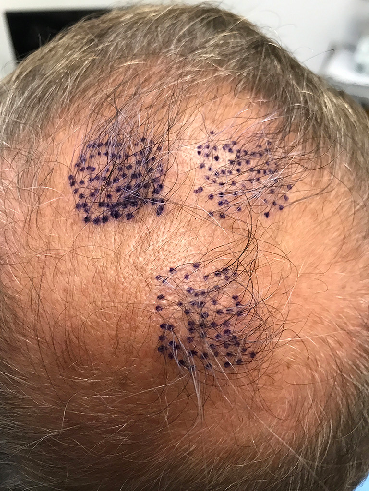
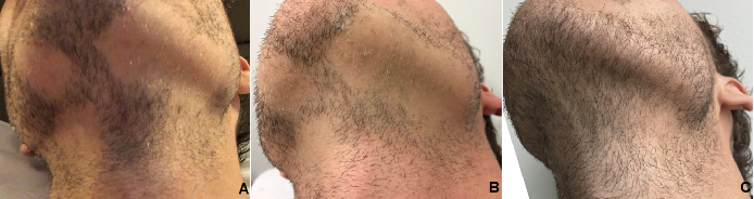
Figure 3 Location of the PL, AA-PRP, and saline treatment zones for Subject C. Forty grafts were placed in each box, and hair checks were conducted 14 weeks post-surgery. PL, …
Alopecia and platelet-derived therapies.
Alopecia and platelet-derived therapies.
Alopecia and platelet-derived therapies.
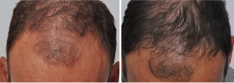
Before
Alopecia and platelet-derived therapies.
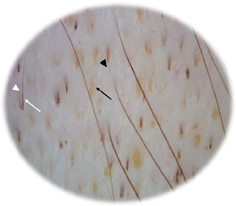
Fig. 1 Representative dermoscopic image from enrolled subjects at baseline showing yellow dots (black arrow), black dots (black triangle), dystrophic hairs (white arrow) and exclamation mark (white triangle)
Efficacy of Postbiotics in a PRP-Like Cosmetic Product for the Treatment of …
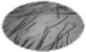
Fig. 2 Representative dermoscopic image from enrolled subjects at baseline (T0)
Efficacy of Postbiotics in a PRP-Like Cosmetic Product for the Treatment of …
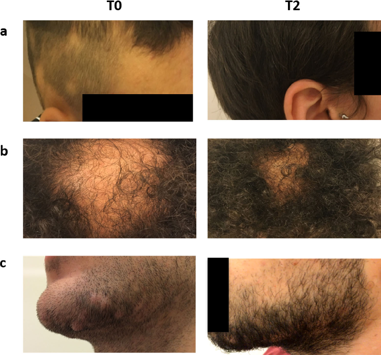
Fig. 3 Digital photographs from three different subjects (a, b, c). Baseline (T0), 3 months of treatment (T2)
Efficacy of Postbiotics in a PRP-Like Cosmetic Product for the Treatment of …
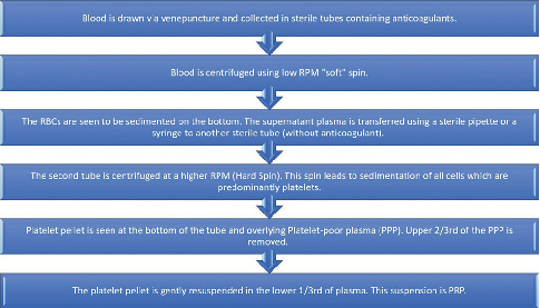
Figure 1: Step-by-step flow diagram of preparation of platelet-rich plasma using open double-spin method
Platelet-Rich Plasma in Androgenetic Alopecia.
Serial photographs document the treatment response to platelet-rich plasma injections, showing minimal regrowth at six weeks after the second injection followed by robust regrowth at one-year follow-up.
Successful Treatment of Alopecia Areata Barbae with Platelet-rich Plasma.
Subgroup analysis of PRP efficacy in alopecia areata stratified by disease severity or PRP preparation technique, revealing potential moderators of treatment response.
Platelet-Rich Plasma in Alopecia Areata-A Steroid-Free Treatment Modality: A Systematic Review and …
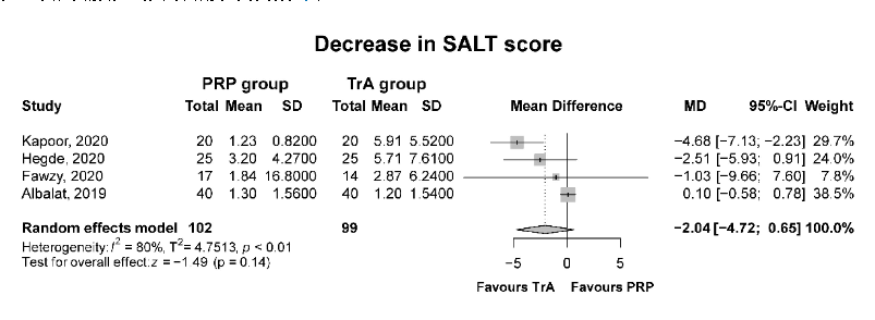
Forest plot depicting the mean decrease in SALT (Severity of Alopecia Tool) score for platelet-rich plasma compared to triamcinolone acetonide, quantifying the relative treatment benefit.
Platelet-Rich Plasma in Alopecia Areata-A Steroid-Free Treatment Modality: A Systematic Review and …
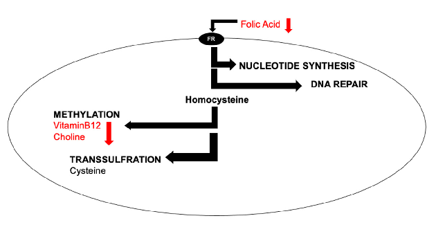
Simplified overview of cellular one-carbon metabolism pathways, illustrating how B-vitamins (folic acid, vitamin B12, choline) participate in nucleotide synthesis, DNA repair, methylation, and transsulfuration reactions relevant to brain health.
The Role of One-Carbon Metabolism in Healthy Brain Aging.
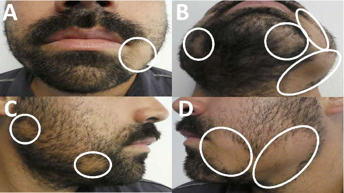
Incipient Diabetes Mellitus and Nascent Thyroid Disease Presenting as Beard Alopecia Areata: …
2페이지 중 1페이지