Spironolactone الأشكال
17 أشكال من أبحاث محكّمة
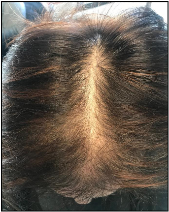
A clinical photograph demonstrating female pattern hair loss, showing characteristic diffuse thinning across the crown and mid-scalp. FPHL affects women with varying degrees of severity and may lead to significant psychological distress.
Female pattern hair loss: A clinical, pathophysiologic, and therapeutic review.
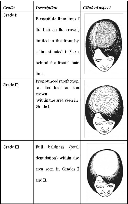
The Ludwig scale classifies female pattern hair loss into three progressive stages, ranging from minimal thinning at the crown (Grade I) to extensive hair loss across the top of the scalp (Grade III). This grading system remains one of the most widely used clinical tools for assessing FPHL severity.
Female pattern hair loss: A clinical, pathophysiologic, and therapeutic review.
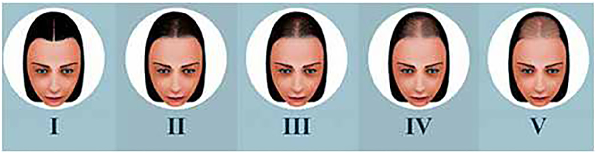
Sinclair's classification divides midline pattern alopecia into four intensity levels, progressing from a normal-appearing scalp to increasingly visible widening of the central part. The scale, introduced by Sinclair et al. (2005), provides a practical visual reference for clinicians assessing hair loss severity.
Female pattern hair loss: A clinical, pathophysiologic, and therapeutic review.
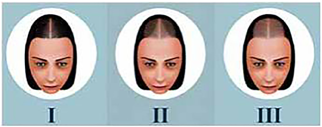
Olsen's classification system highlights the characteristic triangular or Christmas-tree pattern of frontovertical alopecia seen in female pattern hair loss. The accentuation of thinning at the frontal midline distinguishes this pattern from the more diffuse Ludwig classification.
Female pattern hair loss: A clinical, pathophysiologic, and therapeutic review.
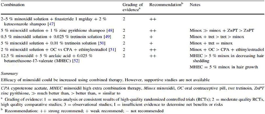
A summary of clinical evidence supporting combination therapy with topical minoxidil for androgenetic alopecia in both men and women. The data, compiled by Varothai and Bergfeld (2014), indicates that multi-modal treatment approaches may offer improved outcomes compared to monotherapy.
Female pattern hair loss: A clinical, pathophysiologic, and therapeutic review.
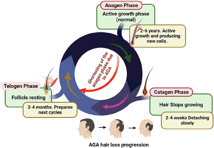
Classification systems for androgenetic alopecia severity are presented, distinguishing male and female pattern hair loss stages.
Androgenetic alopecia: An update.
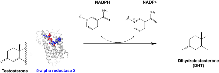
The DHT synthesis pathway via SRD5A2 is diagrammed, showing how 5-alpha reductase converts testosterone to dihydrotestosterone in the presence of NADPH, the key hormonal driver of AGA.
Androgenetic alopecia: An update.
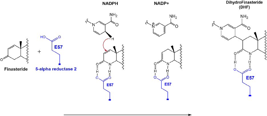
Finasteride's mechanism of inhibition is depicted at the molecular level, showing covalent adduct formation between NADPH and finasteride that prevents further hydride transfer to testosterone.
Androgenetic alopecia: An update.
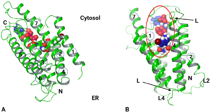
The three-dimensional structure of human SRD5A2 is shown with its seven transmembrane domains, active site, and NADP-DHF adduct positioning within the enzyme channel.
Androgenetic alopecia: An update.
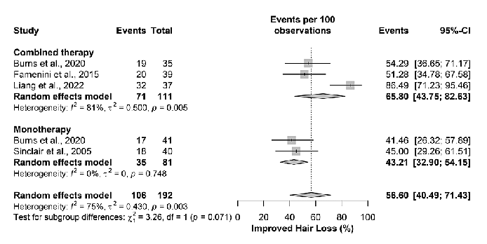
Forest plot or pooled analysis from the spironolactone meta-analysis, presenting combined efficacy data across studies examining oral spironolactone for treating female pattern hair loss.
The Efficacy and Safety of Oral Spironolactone in the Treatment of Female …
Risk of bias or quality assessment for studies included in the spironolactone and female pattern hair loss meta-analysis, evaluating methodological rigor across the evidence base.
The Efficacy and Safety of Oral Spironolactone in the Treatment of Female …
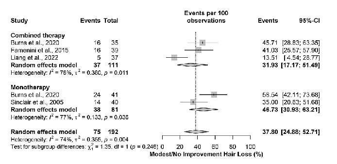
Pooled analysis indicating that hair loss did not improve or showed only modest improvement in 37.80% (95% CI: 24.88-52.71%) of patients treated with oral spironolactone, with subgroup analyses revealing variable response rates.
The Efficacy and Safety of Oral Spironolactone in the Treatment of Female …
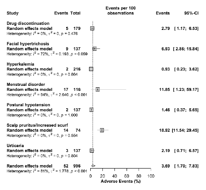
Safety outcomes or adverse event analysis from the spironolactone meta-analysis, summarizing the tolerability profile of oral spironolactone when used for female pattern hair loss.
The Efficacy and Safety of Oral Spironolactone in the Treatment of Female …
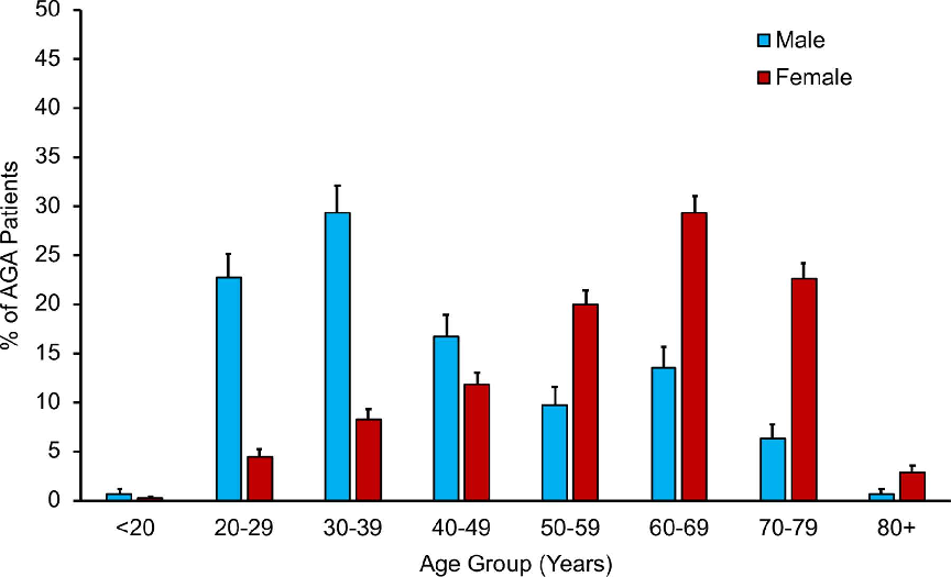
Epidemiological landscape of androgenetic alopecia in the US: An All of Us …
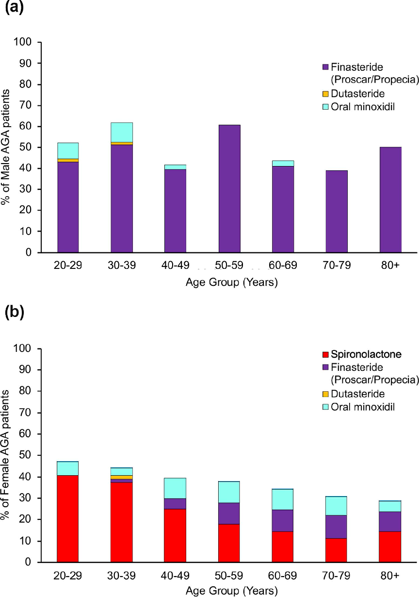
Epidemiological landscape of androgenetic alopecia in the US: An All of Us …
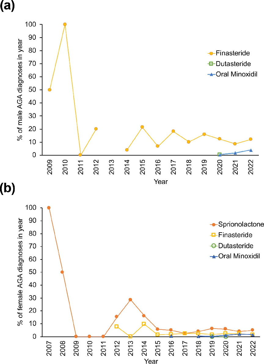
Epidemiological landscape of androgenetic alopecia in the US: An All of Us …
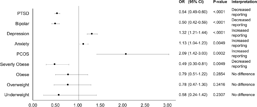
Fig 4. Likelihood of AGA reporting for females by comorbid condition. All data is represented as odds ratios of the condition compared to control with corresponding p-value and interpretation.
Epidemiological landscape of androgenetic alopecia in the US: An All of Us …